Home
/ Microscope Iris Diaphragm Vs Condenser, Kohler Illumination - What is the diaphragm in a compound microscope?
Microscope Iris Diaphragm Vs Condenser, Kohler Illumination - What is the diaphragm in a compound microscope?
Microscope Iris Diaphragm Vs Condenser, Kohler Illumination - What is the diaphragm in a compound microscope?. (1994) contrast in light microscopy: See full list on quekett.org (1998) contrast enhancement techniques for light microscopy in cell biology: The simplest form of condenser is the concave mirror, but this is not useful for objectives above na 0.2 or so. They are all interesting components to consider when focusing your microscope.
See full list on microscopeclarity.com But what happens if our specimen is sensitive to light? Your iris controls the amount of light that enters your cones and rods of your eye by adjusting itself to be larger or smaller. The iris diaphragm is named "iris" mainly because it does the same exact thing as the iris does for our eyes. However, it is difficult to access the back focal plane of the objective (front focal plane when used as a condenser), so supplementary lenses are used to create a position whereby the image of diaphragms and filters are conjugate with the back focal plane.
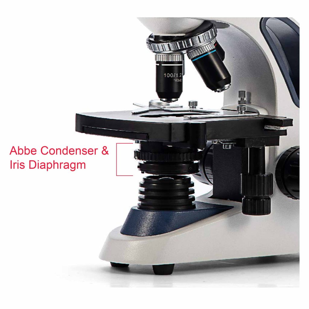
Compound Microscope Basics from optimaxonline.com An example at different settings are below: Here, the ray path is folded about the axis of the specimen where light is reflected from its surface. For example we can use the diaphragm to change how much light will get focused onto the sample. For more on how to focus a microscope see this post. The simplest form of condenser is the concave mirror, but this is not useful for objectives above na 0.2 or so. This is why focusing microscopes can take such a long time. This simple illuminator will suffice for most types of microscopy. Sometimes, the iris diaphragm of a microscope is located within the condenser, in which case it's called an abbe condenser.
It will appear bland and no contrast and almost "blurry".
If your microscope has a mirror and a remote light source, the flat side of the mirror must be used in conjunction with any substage condenser fitted. Adjusting the different kind of diaphragms on a microscope helps the observer to find a good balance between all of them. A less common diaphragm is a disc diaphragm looks a little something like this. Your iris controls the amount of light that enters your cones and rods of your eye by adjusting itself to be larger or smaller. The more common type of diaphragm is the iris diaphragm. Secondly, as mentioned above, it provides a means of regulating contrast (bradbury & evennett, 1996). (1994) contrast in light microscopy: However, the condenser is instrumental in parts (a) and (b) for manipulating contrast and visibility in the image. Sep 12, 2011 · the aperture diaphragm (also called an iris diaphragm) controls contrast, and is found in the condenser, which sits right below the stage in line with the microscope objectives. See full list on quekett.org On the other hand, if you have it almost completely closed, you are preventing a lot of light from getting to the sample. You can never get an image that is high contrast, bright and large. The simplest form of condenser is the concave mirror, but this is not useful for objectives above na 0.2 or so.
See full list on microscopeclarity.com They are all interesting components to consider when focusing your microscope. The first lens converges the incoming light and the second lens focuses the light onto the sample and glass slide (the smiley face). On the other hand, if you have it almost completely closed, you are preventing a lot of light from getting to the sample. Contrast in the image is derived by three means, either separately or in combination.
However, the condenser is instrumental in parts (a) and (b) for manipulating contrast and visibility in the image.
These are a little more sophisticated and are more common among more expensive and more advanced microscopes. The iris diaphragm is named "iris" mainly because it does the same exact thing as the iris does for our eyes. If this is the case for your microscope, you need to find the diaphragm control mechanism on the condenser. Only open the iris diaphragm of the microscope to a point where the light passing through barely extends beyond the microscope's field of view. For example we can use the diaphragm to change how much light will get focused onto the sample. Sep 12, 2011 · the aperture diaphragm (also called an iris diaphragm) controls contrast, and is found in the condenser, which sits right below the stage in line with the microscope objectives. The less light you put in, the more contrast you get. Adjusting the different kind of diaphragms on a microscope helps the observer to find a good balance between all of them. Manipulation of the illumination, and 3. See full list on microscopeclarity.com You need to find the perfect balance between contrast and the total image size and brightness you will get. See full list on quekett.org More images for microscope iris diaphragm vs condenser »
In a case with unhindered light, we have something like this: It will appear bland and no contrast and almost "blurry". Switch it over to the large hole. want less light? These are a little more sophisticated and are more common among more expensive and more advanced microscopes. The condenser fulfills two functions in the microscope.
How To Choose The Right Microscope Compound Microscope Vs Stereo Microscope Rs Science from rsscience.com The microscope diaphragm, also known as the iris diaphragm, controls the amount and shape of the light that travels through the condenser lens and eventually passes through the specimen by expanding and contracting the diaphragm blades that resemble the iris of an eye. Use the condenser diaphragm to reduce the amount of light and increase the contrast of the image. Only open the iris diaphragm of the microscope to a point where the light passing through barely extends beyond the microscope's field of view. Other microscopes have an iris diaphragm with a lever that opens and closes the diaphragm to let in varying amounts of light. This operates in the same way, but this controls how much light and how large the field of view of the resultant image will be. If your microscope has a mirror and a remote light source, the flat side of the mirror must be used in conjunction with any substage condenser fitted. Sep 12, 2011 · the aperture diaphragm (also called an iris diaphragm) controls contrast, and is found in the condenser, which sits right below the stage in line with the microscope objectives. This diaphragm is located closer to the light source of the microscope.
The first lens converges the incoming light and the second lens focuses the light onto the sample and glass slide (the smiley face).
What is the function of the diaphragm on a microscope? However, the condenser is instrumental in parts (a) and (b) for manipulating contrast and visibility in the image. The microscope diaphragm, also known as the iris diaphragm, controls the amount and shape of the light that travels through the condenser lens and eventually passes through the specimen by expanding and contracting the diaphragm blades that resemble the iris of an eye. You need to find the perfect balance between contrast and the total image size and brightness you will get. If this is the case for your microscope, you need to find the diaphragm control mechanism on the condenser. This simple illuminator will suffice for most types of microscopy. Your iris controls the amount of light that enters your cones and rods of your eye by adjusting itself to be larger or smaller. (1994) contrast in light microscopy: Further details of the theoretical and practical aspects of contrast techniques in light microscopy may be found in bradbury & evennett, 1996 and sanderson, 2002, 2000, 1998 and 1994. The condenser may be movable, both in the horizontal and vertical directions. The more common type of diaphragm is the iris diaphragm. Switch it over to the large hole. want less light? The two lenses to the right of the light source are the condenser.
